Thermography

How Does Thermography Work?

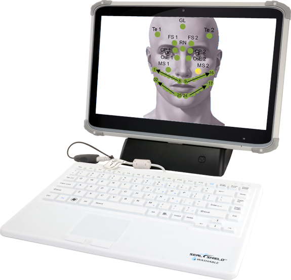
  • Registers skin surface temperature from 112 different points on the body.
  • Displays an image of 25 organs, tissues or systems and their function.
  • Gives a functional perspective based on physiology and cold stress response.
  • Gains a direct index of metabolic activity in various parts of the body.
  • Shows inflammation, degeneration and overactive organs and tissues.

Who Should be Tested?

Anyone over 25 years of age should be tested annually. There is an ever-increasing incidence of asthma, ADD, dermatitis, cancer, chronic fatigue and fibromyalgia. We all know that prevention is best, but how do you prevent what you don’t know is about to happen? Prevention of serious illness is made more realistic via periodic testing and subsequent treatment. The Thermography offers reproducible and scientifically valid information that can be crucial to the development and tracking of a successful treatment strategy.

List of Organs, Tissues and Body Functions Analyzed by Thermography

Breasts, Teeth, Stomach, Liver, Kidneys, Circulation, Gallbladder, Spine, Thyroid, Pancreas, Sinuses, Brain, Sm. Intestine, Tonsils, Lymph, Prostate, Uterus, Ovaries, Colon, Bronchi, and Heart.

Thermography is an FDA-approved, non-invasive diagnostic tool that assesses the function and regulation of the body’s organ systems. This method involves taking temperature readings from over 100 specific points on the surface of the skin, each of which corresponds to underlying organs, glands, or tissues. These readings are taken both before and after exposure to a cold stimulus, allowing practitioners to observe how the body responds to physiological stress. The initial temperatures, along with how quickly and effectively each point recovers from the cold challenge, provide insight into the body’s regulatory capacity and the functional health of various systems.

Unlike structural imaging tools such as X-rays or MRIs, thermography focuses on functional disturbances rather than anatomical changes. This means it can detect early signs of dysregulation or imbalance—often before symptoms appear or more invasive testing would identify an issue. As such, thermography can be an invaluable part of a preventative wellness strategy, revealing potential problem areas that other diagnostic methods may overlook. It is particularly useful for identifying patterns of inflammation, lymphatic congestion, dental and sinus stress, hormonal imbalances, and autonomic nervous system dysfunction.

How does Thermography Use Skin Temperature To Asses Internal Health?

Thermography gathers information by measuring the skin’s surface temperature at numerous specific points that are connected to internal organs, glands, and tissues through the autonomic nervous system. These measurements are taken twice—once at a resting state and again after the body is exposed to a mild cold stimulus, such as cool air. This process evaluates how well the body regulates temperature in response to stress, which in turn reflects the functional state of the organs and systems beneath the skin.

In a healthy response, blood vessels constrict when exposed to cold—a process called vasoconstriction. This reduces blood flow to the skin and helps preserve the body’s core temperature. As a result, the skin temperature at those points should decrease temporarily and then recover within a normal range. However, when an organ or tissue is under stress, inflamed, congested, or not functioning properly, the normal vascular response may be impaired. This results in abnormal temperature patterns—either insufficient cooling, exaggerated cooling, or delayed recovery.

By analyzing the differences between the initial and post-stimulus temperature readings, thermography can detect subtle signs of dysfunction long before structural damage occurs. These temperature patterns can indicate early-stage inflammation, chronic stress, hormonal imbalances, or poor autonomic regulation, providing valuable insight into the overall health and adaptability of the body.

What to Expect

A professional technician will perform the test. You will be asked to sit in a cool room, about 68 degrees. The first measurements of the head, neck, chest and lower abdomen will be taken. This is performed by a gentle touch of the probe to the skin. You will then be asked to remove your clothes, except for your underwear, thereby subjecting your body to the controlled “stress”. You will sit exposed to the air for ten minutes. After this time the measurements are repeated and the test is concluded.

Preparation Instructions

  • Discontinue all non-essential medication 48 hours prior to your test.
  • Do not have any energetic therapy such as acupuncture, a few days prior to the exam.

On the day of your exam

  • Do not smoke.
  • Do not drink coffee or black/green tea.
  • Do not take a hot or cold shower.
  • Do not use cosmetics or lotion on your face or body.
  • Do not exercise the morning of the test.
  • Wear long sleeves and long pants, as loose fitting as possible.
  • Women should not wear a bra.

Videos